

Brain Tutor 3D
Brain Tutor 3D介绍
Explore the brain from the palm of your hand! Learn about the structure and function of the human brain by interacting with high-resolution rotatable 3D models in real-time like you've never experienced it before!
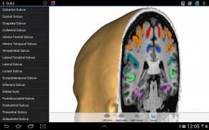
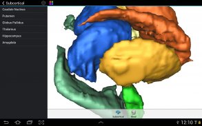
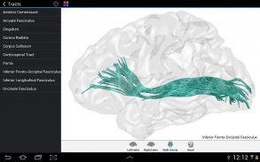
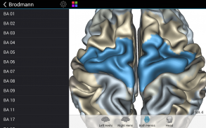
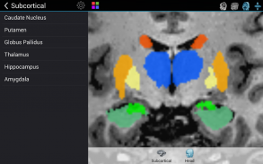
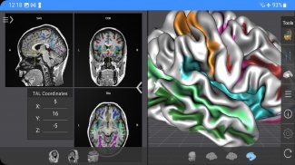
Brain Tutor uses rendered head and brain models as well as fiber tracts that were created from magnetic resonance imaging (MRI) scans of a study volunteer. The MRI data allows to look "inside" the brain using real-time slicing at millimeter resolution. For students, cognitive neuroscientists, medical professionals and everyone interested in the brain, the program provides information about the anatomy and function of the human brain with various atlases describing and visualizing lobes, gyri, sulci, Brodmann areas, subcortical structures, selected specialized functional areas and major fiber tracts.
With Brain Tutor you can:
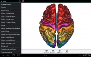
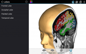
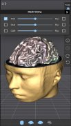
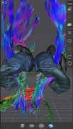
* Explore high-resolution 3D models of the head and brain in real-time.
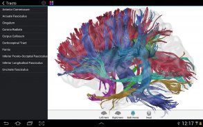
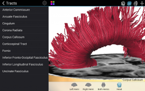
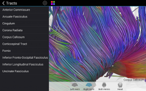
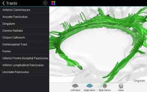
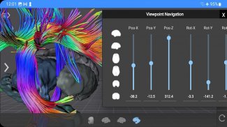
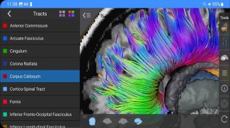
* Visualize major white matter fiber tracts.
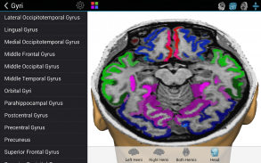
* Slice the brain along three axes (sagittal, axial and coronal).
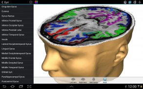
* View MRI brain slices at millimeter resolution.
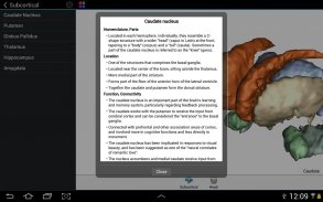
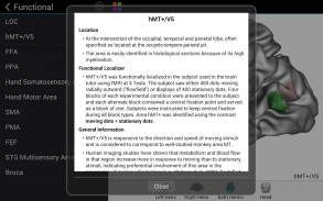
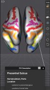
* Learn from text information about the functions of selected lobes, gyri, sulci, subcortical structures, Brodmann areas, functional areas and fiber tracts.
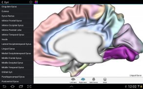
* Learn where brain structures are located both within 3D brain models as well as in MRI slices.
To get started:
* Tap on a 3D brain model to reveal a brain area at that location.
* Switch to another atlas and a specific brain area or fiber tract using the navigation tables.
* Select a 3D model (left/right/both brain hemispheres, head) from the tab bar buttons at the bottom.
* Pan with one finger to rotate a brain model.
* Pan with two fingers to move a brain model.
* Use pinch gesture to zoom a brain model.
* Select the head model to switch to head slicing mode.
* Toggle between navigation and slicing mode by tapping the slicing icon at the top right.
* In head slicing mode, pan with one finger to move the slicing plane through the head.
* Tap on a slicing direction icon in the top bar to switch between three orthogonal slice planes.
* Tap on the displayed name of a selected brain structure to read text information in a popup dialog.
This app has been designed and programmed by Prof. Rainer Goebel, a leading expert in anatomical and functional brain imaging and award-winning developer of scientific software. For more information about his work, see http://www.brainvoyager.com/RainerGoebel.html.
探索大脑从你的手掌!了解人脑通过与高分辨率旋转3D模型的实时交互,就像你从未经历过它的结构和功能!
脑导师使用呈现头部和大脑模型,以及从磁共振成像(MRI)研究志愿者的扫描创建的纤维束。 MRI数据允许使用实时切片在毫米分辨率看“内部”的大脑。对于学生来说,认知神经科学家,医学专家和大家感兴趣的大脑中,程序提供了有关人类大脑的各种地图集描述和可视化叶,脑回,脑沟,布罗德曼区,皮层下结构,选择的专业功能区的解剖和功能信息和主要纤维束。
脑导师,您可以:
探索在实时的头部和脑部的高分辨率三维模型。
*可视化主要白质纤维束。
*切片沿三个轴(矢状位,轴位和冠状)的大脑。
*查看核磁共振脑部切片在毫米分辨率。
*从大约选中叶,脑回,脑沟,皮层下结构,布罗德曼区,功能区和纤维束的功能文字信息了解。
*了解那里的大脑结构位于两个内3D脑模型,以及在MRI片。
上手:
*点选一个3D的大脑模型,揭示了大脑区域在该位置。
*切换到另一个地图集和特定脑区或纤维束使用导航表。
*从底部的标签栏按钮选择3D模型(左/右/两个大脑半球,头)。
*用一个手指平移旋转的大脑模型。
*用两个手指平移来移动一个大脑模型。
*使用捏的手势来放大大脑模型。
*选择头部模型切换到头部切片模式。
*通过点击切片图标右上角的导航和切片模式之间切换。
*在头切片模式,泛用一根手指穿过头部移动的切割平面。
*轻按在上面扎上切片的方向图标,三个正交切片平面之间切换。
*轻触选定的大脑结构的显示名称来读取文本信息在弹出的对话框中。
这个应用程序的设计和教授莱纳格贝尔,在解剖和功能性脑成像和屡获殊荣的科学软件开发的领先专家编程。有关他的工作的更多信息,请参阅http://www.brainvoyager.com/RainerGoebel.html。
Brain Tutor 3D - 版本2.0
(27-02-2023)Brain Tutor 3D - APK信息
APK版本: 2.0程序包: com.brainvoyager.android.BrainTutorBrain Tutor 3D的最新版本
其他版本


同类应用